Distal Biceps Tendon Rupture Repair: A Comparative Analysis of Surgical Techniques and Postoperative Management
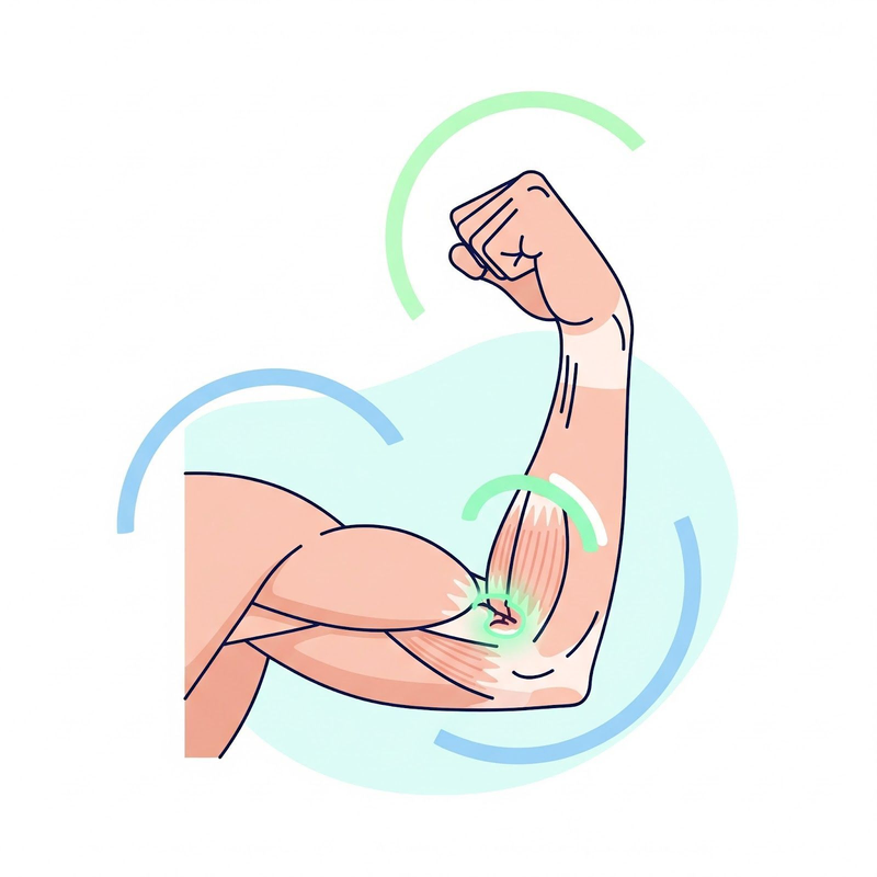
Ruptures of the distal biceps tendon are often characterized by a sudden "pop" in the elbow, followed by pain, bruising, and weakness in bending the elbow or twisting the forearm 1. This type of injury commonly occurs in active individuals, especially men aged 30 to 50, when the tendon connecting the biceps muscle to the radius bone in the forearm tears due to forceful contraction or an eccentric load on a flexed elbow 2. If left untreated, these ruptures can lead to a significant decrease in supination (rotating the forearm palm up) and flexion (bending the elbow) strength 4. It is important to note that surgical repair is most successful when performed within the first four weeks following the injury. After this period, the tendon and biceps muscle begin to scar and shorten, making it more challenging to restore full arm function 5.
This article provides a comprehensive overview of distal biceps tendon rupture repair, comparing and contrasting different surgical techniques and highlighting the crucial role of early mobilization and rehabilitation in optimizing patient outcomes.
Surgical Techniques for Distal Biceps Tendon Repair
The two primary surgical approaches for repairing distal biceps tendon ruptures are the single-incision and double-incision techniques. Both methods aim to reattach the torn tendon to the radial tuberosity, but they differ in their approach and potential complications. A systematic review and meta-analysis of comparative studies found no significant differences in functional scores between the single-incision and double-incision approaches 6. However, the single-incision approach showed greater range of motion in flexion and pronation and a lower risk of heterotopic ossification and reoperation, while the double-incision approach was associated with a lower risk of neurological complications 6.
Single-Incision Technique
The single-incision technique involves a single anterior incision in the antecubital fossa (the crease of the elbow) 4. This approach allows the surgeon to access and repair the tendon with minimal disruption to surrounding tissues. In one study, the single-incision technique utilized two suture anchors for tendon fixation 4. The Endobutton implant is another example of a fixation device used in this approach 7. Advantages of the single-incision technique include:
- Reduced risk of heterotopic ossification: Heterotopic ossification is the formation of bone in soft tissues where bone normally does not exist. The single-incision technique has been associated with a lower incidence of heterotopic ossification compared to the double-incision approach 2.
- Faster recovery: With less surgical dissection, patients undergoing the single-incision technique may experience faster wound healing and a quicker return to activity 8.
- Improved cosmetic outcome: A single incision generally results in a smaller scar compared to two incisions.
- Greater range of motion: Studies have shown that the single-incision approach results in greater range of motion in flexion and pronation compared to the double-incision technique 6.
However, the single-incision technique also has potential drawbacks:
- Increased risk of nerve injury: The lateral antebrachial cutaneous nerve, which provides sensation to the forearm, is at risk of injury during the single-incision approach. Studies have reported a higher incidence of nerve complications, such as neurapraxia (temporary nerve dysfunction) and paresthesia (numbness or tingling), with the single-incision technique 2.
- Challenges with anatomical tendon placement: Some studies suggest that achieving precise anatomical reattachment of the tendon to the radial tuberosity can be more challenging with the single-incision technique 9. This may potentially affect long-term outcomes in terms of strength and stability.
- Higher rerupture rate: Single-incision repairs have a greater rate of failed reattachment and rerupture compared to double-incision repairs 2.
Double-Incision Technique
The double-incision technique utilizes two separate incisions: one in the antecubital fossa and another on the posterolateral side of the elbow 8. This approach provides better visualization of the radial tuberosity and allows for more anatomical tendon placement. Advantages of the double-incision technique include:
- Reduced risk of nerve injury: By avoiding extensive dissection in the antecubital fossa, the double-incision technique minimizes the risk of lateral antebrachial cutaneous nerve injury 6.
- Improved anatomical tendon placement: The double-incision approach facilitates more accurate reattachment of the tendon to its original insertion site on the radial tuberosity, potentially leading to better strength and stability 9.
However, the double-incision technique also has potential disadvantages:
- Increased risk of heterotopic ossification: The more extensive surgical dissection involved in the double-incision technique has been associated with a higher incidence of heterotopic ossification 2.
- Longer recovery time: With two incisions, patients may experience a slightly longer recovery period compared to the single-incision technique.
- Increased risk of radioulnar synostosis: Radioulnar synostosis is an abnormal bony connection between the radius and ulna bones in the forearm 10. While rare, this complication has been reported more frequently with the double-incision technique 10.
Choosing the Optimal Surgical Approach
The choice between single-incision and double-incision techniques for distal biceps tendon repair depends on various factors, including the surgeon's experience, the patient's individual needs, and the specific characteristics of the injury. Surgeons carefully consider the potential risks and benefits of each approach to determine the most appropriate technique for each patient. For example, a patient with obesity may be a better candidate for a double-incision approach due to the increased risk of complications with the single-incision technique in this population 9. Additionally, the timing of the repair can influence the choice of surgical approach. Patients presenting in a delayed fashion may experience a higher rate of transient neurapraxia after surgical repair, although final functional outcomes have not been shown to differ compared to those of acute repairs 9.
| Technique | Advantages | Disadvantages |
|---|---|---|
| Single-Incision | Reduced risk of heterotopic ossification, faster recovery, improved cosmetic outcome, greater range of motion | Increased risk of nerve injury, challenges with anatomical tendon placement, higher rerupture rate |
| Double-Incision | Reduced risk of nerve injury, improved anatomical tendon placement | Increased risk of heterotopic ossification, longer recovery time, increased risk of radioulnar synostosis |
Importance of Early Mobilization and Rehabilitation
Regardless of the surgical technique used, early mobilization and rehabilitation are crucial for optimizing outcomes after distal biceps tendon repair. Early mobilization involves gentle movement of the elbow soon after surgery, while rehabilitation encompasses a structured program of exercises and activities to restore strength, range of motion, and function.
Early mobilization and rehabilitation are essential for several reasons. First, movement promotes blood circulation to the injured area, which helps deliver nutrients and oxygen essential for tissue healing and reduces postoperative pain and swelling 11. Second, early mobilization helps prevent the formation of excessive scar tissue, which can restrict movement and lead to long-term stiffness 12. Third, gentle movement helps maintain joint flexibility and facilitates a quicker return to normal range of motion 13. Finally, studies have shown that early mobilization can lead to greater patient satisfaction with the overall outcome of surgery 13.
Rehabilitation Protocols
Rehabilitation after distal biceps tendon repair typically involves a phased approach, with exercises and activities gradually progressing in intensity as the tendon heals. Common rehabilitation protocols include:
- Phase 1 (Weeks 1-2): This phase focuses on protecting the repair, decreasing pain and inflammation, and initiating early motion. Patients may be placed in a brace and instructed to perform passive range of motion exercises for the elbow, hand, and wrist, as well as scapular exercises 14.
- Phase 2 (Weeks 3-6): In this phase, active range of motion is gradually increased, and light isotonic exercises are introduced. Patients may begin performing gravity-assisted flexion and extension exercises, as well as isometric triceps exercises 14.
- Phase 3 (Weeks 7-12): This phase focuses on continued improvement in range of motion and strengthening. Isotonic triceps exercises and strengthening of wrist flexors and extensors are initiated. Patients may also begin performing light isotonic biceps exercises 14.
- Phase 4 (Weeks 12+): This phase emphasizes advanced strengthening and a gradual return to activity. Patients may begin performing biceps curls with varying grips, triceps extensions, and rotator cuff and periscapular strengthening exercises 14.
Specific exercises commonly incorporated into rehabilitation protocols include:
- Passive range of motion exercises: Gentle movements of the elbow and forearm assisted by the therapist or the patient's other hand 14. Active extension, where the patient pushes their elbow straight, and passive flexion, where the patient uses their uninjured hand to pull the injured arm back, are examples of these exercises 17.
- Active range of motion exercises: Unassisted movements of the elbow and forearm 17.
- Strengthening exercises: Exercises using resistance bands, weights, or bodyweight to strengthen the biceps, triceps, and other muscles around the elbow 14.
- Functional exercises: Activities that mimic daily tasks or sports movements 11.
Risks of Delaying Mobilization and Rehabilitation
Delaying mobilization and rehabilitation can have several adverse effects, including:
- Increased stiffness and limited range of motion: Prolonged immobilization can lead to joint stiffness and difficulty regaining full range of motion 19.
- Muscle weakness and atrophy: Lack of use can cause the muscles around the elbow to weaken and shrink, further delaying functional recovery 21.
- Increased risk of complications: Delayed mobilization may increase the risk of complications such as heterotopic ossification and nerve injury 9.
- Prolonged recovery time: Overall, delaying mobilization and rehabilitation can significantly extend the recovery period and delay the return to normal activities. For example, one study found that delaying repair beyond 3 weeks may result in increased exertional pain and paresthesia of the lateral antebrachial cutaneous nerve 22.
Conclusion
Distal biceps tendon rupture repair is a crucial intervention for restoring function and preventing long-term disability in active individuals. Both single-incision and double-incision techniques have been shown to be effective, with each approach having its own advantages and disadvantages. The choice of surgical technique depends on various factors, including patient characteristics, timing of the repair, and surgeon experience. Surgeons carefully consider the patient's individual needs and the specific characteristics of the injury to determine the most appropriate technique.
Regardless of the surgical approach, early mobilization and a comprehensive rehabilitation program are essential for optimizing outcomes. Early mobilization promotes healing, reduces stiffness, and facilitates a faster recovery of range of motion. Rehabilitation protocols typically involve a phased approach, with exercises and activities gradually progressing in intensity as the tendon heals. Delaying mobilization and rehabilitation can have adverse effects, including increased stiffness, muscle weakness, and prolonged recovery time.
Ultimately, successful outcomes after distal biceps tendon repair rely on individualized surgical decision-making and a commitment to early mobilization and diligent participation in a comprehensive rehabilitation program.
Works cited
1. Distal Biceps Tendon Repair - Orthosports Info, accessed February 17, 2025, https://www.orthosports.info/distal-biceps-tendon-repair/
2. www.drronakpatel.com, accessed February 17, 2025, /images/press/uploads/2023/01/11-Complications-of-Distal-Biceps-Tendon-Repair.pdf
3. Complications of Distal Biceps Tendon Repair: A Meta-analysis of ..., accessed February 17, 2025, https://pmc.ncbi.nlm.nih.gov/articles/PMC5056595/
4. upload.orthobullets.com, accessed February 17, 2025, https://upload.orthobullets.com/journalclub/free_pdf/22760383_DistalBiceps-JBJS2013-El-Hawary.pdf
5. Distal Biceps Rupture Surgery - Dr David Duckworth, accessed February 17, 2025, https://www.drdavidduckworth.com.au/patient-information/biceps-rupture-surgery/
6. Single- versus double-incision technique for the treatment of distal ..., accessed February 17, 2025, https://boneandjoint.org.uk/Article/10.1302/0301-620X.102B12.BJJ-2020-0822.R2
7. cdn-uat.mdedge.com, accessed February 17, 2025, https://cdn-uat.mdedge.com/files/s3fs-public/Document/September-2017/ajo04307E159.pdf
8. Comparative Analysis of Surgical Approaches for Distal Biceps ..., accessed February 17, 2025, https://www.mdpi.com/2077-0383/12/19/6423
9. Distal Biceps Tendon Rupture: JBJS Clinical Summary, accessed February 17, 2025, https://www.jbjs.org/summary.php?id=220
10. Major complications after distal biceps tendon repairs: retrospective cohort analysis of 970 cases - Orthobullets, accessed February 17, 2025, https://upload.orthobullets.com/journalclub/free_pdf/30139681_30139681.pdf
11. distal biceps tendon repair protocol - Mark Adickes, M.D., accessed February 17, 2025, https://www.jocktodoc.com/pdf/distal-biceps-tendon-repair-new.pdf
12. Rehabilitation And Recovery After Distal Biceps Tendon Repair, accessed February 17, 2025, https://sydneyorthopaedicsurgeon.com.au/rehabilitation-and-recovery-after-distal-biceps-tendon-repair/
13. Does immediate elbow mobilization after distal biceps tendon repair carry the risk of wound breakdown, failure of repair, or patient dissatisfaction? - PubMed, accessed February 17, 2025, https://pubmed.ncbi.nlm.nih.gov/26897313/
14. REHABILITATION FOLLOWING DISTAL BICEPS REPAIR - PMC, accessed February 17, 2025, https://pmc.ncbi.nlm.nih.gov/articles/PMC6449020/
15. Distal Biceps Repair Clinic Practice Guideline - Ohio State College of Medicine, accessed February 17, 2025, https://medicine.osu.edu/-/media/files/medicine/departments/sports-medicine/medical-professionals/shoulder-and-elbow/distal-biceps-repair2021.pdf?la=en&hash=0C6FE7E59999DF0D6D69AF9C53423AF8872A1B52
16. distal-biceps-tendon-repair-protocol.pdf - Dr Katherine Coyner, accessed February 17, 2025, https://www.drcoyner.com/pdf/distal-biceps-tendon-repair-protocol.pdf
17. Distal biceps Tendon Repair Initial Postoperative Stretching Exercises - YouTube, accessed February 17, 2025,
18. Distal Biceps Repair Protocol - The Royal Melbourne Hospital, accessed February 17, 2025, https://www.thermh.org.au/files/documents/Patients/Fracture/distal-bicep-repair-protocol.pdf
19. Distal Biceps Tendon Rupture - Physiopedia, accessed February 17, 2025, https://www.physio-pedia.com/Distal_Biceps_Tendon_Rupture?utm_source=physiopedia&utm_medium=related_articles&utm_campaign=ongoing_internal
20. Distal biceps repair - Windsor Upper Limb, accessed February 17, 2025, https://www.windsorupperlimb.com/procedures/elbow-procedures/distal-biceps-repair
21. REHABILITATION FOLLOWING DISTAL BICEPS REPAIR | Matthew Provencher, MD, accessed February 17, 2025, /images/press/uploads/2021/03/REHABILITATION-FOLLOWING-DISTAL-BICEPS-REPAIR.pdf
22. Clinical outcomes after refixation of subacute repaired distal biceps tendon ruptures - PMC, accessed February 17, 2025, https://pmc.ncbi.nlm.nih.gov/articles/PMC9091753/