Distal Humerus Fractures: Surgical Management and Post-Operative Rehabilitation
Distal humerus fractures, breaks in the lower end of the upper arm bone (humerus), are a complex injury that can significantly impact elbow joint function. These fractures represent approximately 2% of all fractures and 30% of elbow fractures, with a rising incidence. They are most often caused by falls directly on the elbow, high-energy impacts in younger populations (such as those sustained during motor vehicle collisions), and falls on an outstretched arm with the elbow held tightly to brace against the fall1. This comprehensive review delves into the surgical management of these fractures, focusing on different surgical approaches and post-operative rehabilitation protocols. Additionally, it explores potential complications and factors influencing treatment decisions.
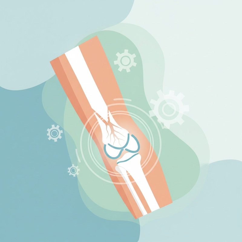
Anatomy of the Distal Humerus
Understanding the anatomy of the distal humerus is crucial for comprehending the complexity of these fractures and their surgical management. The distal humerus can be conceptualized as two columns, medial and lateral, each with a corresponding epicondyle1. The medial and lateral columns form a triangular shape. The articular surface of the distal humerus, where it meets the bones of the forearm (radius and ulna), is composed of the capitellum and trochlea1. Important ligaments, such as the medial collateral ligament (MCL) and lateral collateral ligament (LCL), provide stability to the elbow joint1.
Diagnosis
Diagnosing a distal humerus fracture typically involves a combination of physical examination and imaging studies1. During the physical exam, the physician will assess the elbow for pain, swelling, tenderness, and any signs of open wounds. They will also evaluate the range of motion and stability of the joint and conduct a thorough neurovascular examination to check for any nerve or blood vessel damage1. Imaging studies, such as X-rays, are essential to confirm the diagnosis and determine the type and extent of the fracture1. In some cases, a CT scan may be necessary to provide a more detailed view of the fracture, especially if there is intra-articular involvement1.
Surgical Management of Distal Humerus Fractures
Surgical intervention is often necessary for distal humerus fractures, especially when the bones are displaced or break through the skin (open fracture)2. The primary goals of surgery are to restore anatomical alignment, achieve stable fixation, and enable early mobilization to minimize complications and optimize functional outcomes3.
Types of Distal Humerus Fractures
The AO/OTA classification system is commonly used to categorize distal humerus fractures based on their complexity and involvement of the articular surface: 1
- Type A: Extra-articular: These fractures occur above the elbow joint and do not involve the articular surface. They are often supracondylar fractures, with 80% being extension type.
- Type B: Partial Articular: These fractures involve a single column of the distal humerus and extend into the articular surface. They can be further classified as isolated condylar, coronal shear, or epicondyle fractures with articular extension.
- Type C: Complete Articular: These fractures involve both columns of the distal humerus, and no part of the joint remains connected to the shaft. They represent complete articular fractures.
Each type is further subdivided based on the degree and location of fracture comminution.
Surgical Approaches
Various surgical approaches can be employed to access and repair distal humerus fractures. The choice of approach depends on factors such as fracture complexity, surgeon preference, and the need for exposure of specific anatomical structures4. Common approaches include:
-
Posterior Approach: This is the most common approach, providing good visualization of the articular surface and fracture fragments4. It involves an incision along the back of the elbow, with variations in how the triceps muscle is managed. The patient is positioned laterally with the arm over an "L" holder3. A midline posterior incision is made, curving down lateral to the olecranon3. Full-thickness medial and lateral flaps are created3. If the dissection is extended proximally, the radial nerve should be identified and protected3. A capsulotomy is performed on either side of the olecranon3.
-
Triceps-Sparing Approach: The triceps muscle is carefully retracted to preserve its function and minimize complications. This approach is suitable for less complex fractures5.
- Triceps-Reflecting Approach: The triceps muscle is detached and later reattached, offering better exposure for complex fractures5.
- Olecranon Osteotomy: The olecranon process (bony tip of the elbow) is temporarily cut to allow wider access to the joint. This provides excellent visualization but carries a higher risk of complications like nonunion or hardware prominence4. An apex distal chevron osteotomy is performed with an oscillating saw and completed with an osteotome3.
-
Lateral Approach: Used for isolated single column or epicondylar injuries7.
-
Medial Approach: Also used for isolated single column or epicondylar injuries7.
-
Combined Medial-Lateral Approach: Employed for some intra-articular fractures7.
Surgical Techniques
The most common surgical technique for distal humerus fractures is Open Reduction and Internal Fixation (ORIF) 2. This involves:
- Making an incision to access the fracture site.
- Reducing the fracture, which means putting the bone fragments back into their correct anatomical position.
- Fixating the fracture using implants such as metal plates, screws, wires, or pins to hold the bones in place while they heal8.
ORIF allows for optimal reduction and direct access to reduce fracture fragments with advanced devices9. It also facilitates early motion, which is crucial for preventing stiffness and achieving optimal functional outcomes9.
In some cases, fixation may be achieved solely with Kirschner wires or screws3. However, plate fixation is generally preferred as it provides more rigid and stable fixation, leading to better functional outcomes3.
In cases of severe comminution (bone broken into many pieces) or significant bone loss, an external fixator may be used as a temporary stabilization method2. This involves placing pins into the bone above and below the fracture, with the pins connected to an external frame.
For some complex fractures, particularly in elderly patients with severely damaged bone, total elbow replacement (arthroplasty) may be considered2. This involves replacing the damaged portions of the humerus and ulna with artificial components.
Fixation Principles
Regardless of the specific approach or technique used, certain principles guide the internal fixation of distal humerus fractures: 7
- Ensure every screw passes through a plate and engages a fragment on the opposite side, also fixed to a plate.
- Maximize the number of screws placed in the distal fragments.
- Use screws that are as long as possible to engage the opposite cortex.
- Engage as many articular fragments as possible with each screw.
- Achieve compression at the supracondylar level for both columns.
- Use plates strong enough to resist breaking or bending before the bone heals.
Factors Influencing Surgical Approach
Several factors influence the choice of surgical approach for distal humerus fractures: 7
- Fracture Type: The complexity and location of the fracture, including the degree of articular involvement and comminution, play a significant role in determining the approach.
- Bone Quality: In patients with osteoporosis or poor bone quality, more stable fixation constructs may be necessary, influencing the choice of approach.
- Soft Tissue Injury: The extent of any associated soft tissue injury may influence the incision placement and approach.
- Surgeon Experience and Preference: Surgeons may have preferences for certain approaches based on their training and experience.
- Patient Factors: The patient's age, overall health, and functional demands are also considered.
For example, in extra-articular fractures, a standard paratricipital approach with medial and lateral windows may be sufficient4. For simple intra-articular fractures, an anconeus flap can be created for better exposure4. In more complex intra-articular fractures, an olecranon osteotomy may be necessary to achieve adequate visualization3.
Post-Operative Rehabilitation Protocols
Post-operative rehabilitation is crucial for restoring elbow function, range of motion, and strength after distal humerus fracture surgery. Rehabilitation protocols are typically tailored to the individual patient, considering factors such as fracture type, surgical approach, and any associated injuries7. However, general guidelines and stages can be outlined:
| Phase | Timeframe | Immobilization | Range of Motion | Weight-Bearing | Strengthening | Other Interventions |
|---|---|---|---|---|---|---|
| Phase I: Early Motion and Protection | Weeks 1-4 | Splint or brace worn at all times for 1 week, then only when outside for 3 more weeks10. | Passive flexion and abduction to 90°, external rotation as tolerated at 0° abduction, internal rotation as tolerated10. | Non-weight-bearing (NWB)10. | Cervical ROM exercises; elbow, wrist, hand ROM; active elbow flexion/extension; shoulder shrugs and scapula retraction; wall walking or table slides for flexion and abduction (all as tolerated)10. | Pain management with medications and ice11. |
| Phase II: Advanced Motion and Function | Weeks 4-6 | Discontinue sling at week 4, but can be worn for comfort10. | As tolerated in all planes. No aggressive or forced passive ROM until full union of fracture10. | NWB10. | Begin no-load serratus exercises at 5-6 weeks; begin limited range, no resisted active external rotation/internal rotation with towel roll; submaximal isometrics as tolerated10. | |
| Phase III: Full Motion and Function | Weeks 6-9 | Not needed10. | As tolerated in all planes10. | Partial weight-bearing (PWB)10. | Begin posterior capsule stretches; begin upper body exercises below shoulder level; progressive scapular strengthening; begin rows with theraband; begin light band theraband resistance exercises in all planes10. | |
| Phase IV: Return to Activity | Weeks 9-12 | Not needed10. | Continue working towards full active range of motion (FAROM)10. | PWB10. | Increase resistance with theraband exercises; advance kinesthetic awareness exercise to multi-angle; closed kinetic chain progression of upper extremity muscles10. | |
| Phase V: Full Recovery | Months 3-6 | Not needed10. | FAROM10. | Weight-bearing as tolerated (WBAT)10. | Advance strengthening for rotator cuff and rest of the upper extremity; advance scapular exercises; no overhead lifting for 4-6 months post-op; sport-specific training10. |
It is important to emphasize that early mobilization is a key principle in post-operative rehabilitation3. Starting gentle range of motion exercises soon after surgery helps prevent stiffness, promotes healing, and optimizes functional outcomes.
Factors Influencing Rehabilitation Protocol
Several factors can influence the specific rehabilitation protocol for a patient with a distal humerus fracture: 7
- Fracture Type: The type and severity of the fracture, including the degree of comminution and displacement, will affect the rehabilitation plan.
- Surgical Approach: The surgical approach used, such as whether an olecranon osteotomy was performed, will influence the initial phases of rehabilitation.
- Fixation Stability: The stability of the fixation achieved during surgery will determine the allowable range of motion and weight-bearing restrictions.
- Individual Patient Factors: The patient's age, overall health, and functional demands will be considered when tailoring the rehabilitation program.
- Associated Injuries: Any associated injuries, such as nerve damage or ligament injuries, will need to be addressed in the rehabilitation plan.
For example, medial epicondylar fractures are immobilized for 7 days with the elbow flexed at 90°, the forearm pronated, and the wrist flexed at 30° to relax the muscles7.
Potential Complications
While surgical management and rehabilitation generally yield good outcomes, potential complications can arise: 2
| Complication | Description | Impact on Rehabilitation |
|---|---|---|
| Elbow Stiffness | Loss of motion in the elbow joint, often due to scar tissue formation or prolonged immobilization1. | May require intensive physical therapy, special bracing, or further surgery to regain motion2. |
| Heterotopic Ossification | Formation of bone in the soft tissues around the elbow joint1. | Can limit range of motion and require surgical removal if severe2. |
| Nonunion | Failure of the fracture to heal properly1. | May necessitate revision surgery with bone grafting1. |
| Malunion | Healing of the fracture in a non-anatomical position1. | Can lead to long-term functional limitations and may require corrective surgery. |
| Infection | Infection at the surgical site2. | May delay healing and require antibiotics or further surgery. |
| Nerve Injury | Damage to nerves around the elbow, such as the ulnar nerve or radial nerve2. | Can cause numbness, weakness, or pain and may require further surgery2. |
| Blood Vessel Damage | Injury to blood vessels around the elbow12. | Can compromise blood supply to the arm and hand and may require vascular surgery. |
| Hardware Complications | Problems with the implanted hardware, such as loosening, breakage, or migration12. | May necessitate revision surgery. |
| Anesthetic Complications | Adverse reactions to anesthesia12. | Can range from minor side effects to serious complications. |
| Posttraumatic Arthritis | Development of arthritis in the elbow joint due to cartilage damage2. | Can cause pain and stiffness and may require further surgery in severe cases2. |
Conclusion
Distal humerus fractures present a complex challenge in orthopedic surgery. Achieving optimal outcomes requires a comprehensive approach that includes careful preoperative planning, appropriate surgical technique, and a well-structured rehabilitation program. This review has highlighted the various surgical approaches, techniques, and rehabilitation protocols for managing these fractures. It has also emphasized the importance of considering individual patient factors, such as age, bone quality, and functional demands, when making treatment decisions. While surgical management and rehabilitation generally yield good outcomes, potential complications can arise. Early recognition and appropriate management of these complications are crucial for minimizing their impact on long-term function. By staying informed about the latest advancements and best practices in the field, medical professionals can provide the best possible care for patients with distal humerus fractures.
Works cited
1. Distal Humerus Fractures - Trauma - Orthobullets, accessed February 17, 2025, https://www.orthobullets.com/trauma/1017/distal-humerus-fractures
2. Distal Humerus Fractures of the Elbow - OrthoInfo - AAOS, accessed February 17, 2025, https://orthoinfo.aaos.org/en/diseases--conditions/distal-humerus-fractures-of-the-elbow/
3. Distal Humeral Fractures-Current Concepts - The Open Orthopaedics Journal, accessed February 17, 2025, https://openorthopaedicsjournal.com/VOLUME/11/PAGE/1353/FULLTEXT/
4. Distal humerus fractures: review of literature, tips, and tricks - PMC - PubMed Central, accessed February 17, 2025, https://pmc.ncbi.nlm.nih.gov/articles/PMC11329030/
5. Distal humerus fractures: a review of current therapy concepts - PMC, accessed February 17, 2025, https://pmc.ncbi.nlm.nih.gov/articles/PMC4896884/
6. Distal Humerus Fractures - StatPearls - NCBI Bookshelf, accessed February 17, 2025, https://www.ncbi.nlm.nih.gov/books/NBK531474/
7. Distal Humerus Fractures Treatment & Management - Medscape Reference, accessed February 17, 2025, https://emedicine.medscape.com/article/1239515-treatment
8. Distal Humerus Fracture Treatment Salt Lake City | Elbow Deformity ..., accessed February 17, 2025, https://www.roberttashjianmd.com/humerus-fracture-orthopaedics-surgeon-salt-lake-city-ut.html
9. Open Reduction & Internal Fixation of Proximal Humerus Fracture - Dr Mauricio Herrera, accessed February 17, 2025, https://www.herrerasportsmedicine.com/open-reduction-internal-fixation-of-proximal-humerus-fracture-miami-institute.html
10. Distal Humerus Fracture Rehab Protocol | Ortho.Boston, accessed February 17, 2025, https://www.ortho.boston/distal-humerus-fracture-rehab
11. Elbow, Radial Head, Distal Humerus, Coronoid Fractures, accessed February 17, 2025, /images/press/uploads/2016/06/Elbow-Radial-Head-Distal-Humerus-Coronoid-Fractures.pdf
12. ORIF of the Distal Humerus Fractures - Dr Clayton Nelson, accessed February 17, 2025, https://www.oklahomahand.com/orif-distal-humerus-fractures-upper-extremity-surgeon-edmond-norman-oklahoma.html