Beyond X-Rays: A Look at Special Imaging for Musculoskeletal Health
When it comes to musculoskeletal health, X-rays are often the first step. But what happens when doctors need a more detailed view? That's where special imaging studies come in. From the power of MRI to the precision of CT scans, these tools offer a deeper look at bones, joints, and soft tissues.
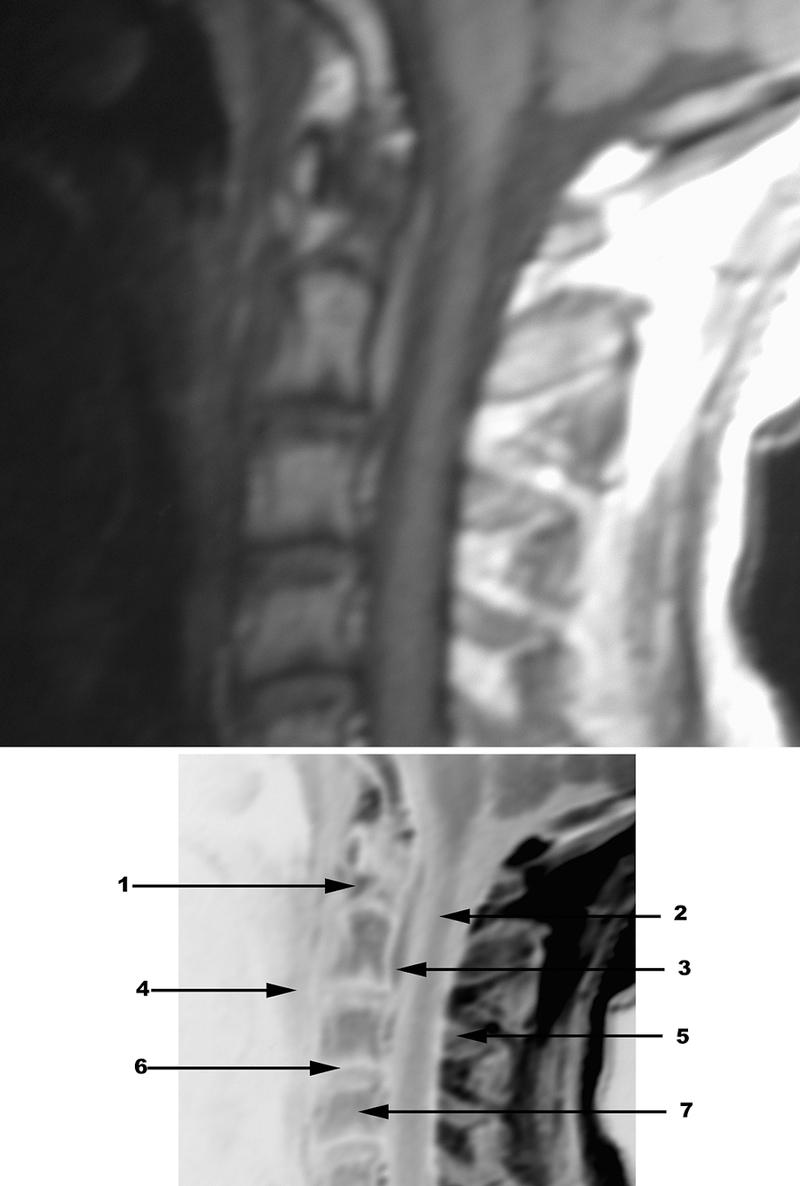
Think of an MRI as a high-definition camera for your insides. It's fantastic for spotting subtle changes in bone marrow, cartilage, and ligaments. Got a sports injury? An MRI can help pinpoint those tricky tears. While it's a bit pricey and can't play well with metal implants, its detailed images are invaluable.
Then there's the CT scan, the master of bone detail. If doctors need to see calcifications, cortical bone changes, or complex fractures, CT is their go-to. It can even create 3D models, which are super helpful for surgical planning.
Nuclear medicine bone scans are like detectives. They use radioactive tracers to find areas of increased bone activity, which can signal tumors, infections, or fractures. It's a very sensitive test, though it might not always tell the whole story on its own.
Arthrography and MR Arthrography take imaging a step further by injecting contrast material into joints. This helps visualize joint structures and spot issues like rotator cuff tears or meniscal problems.
And let's not forget ultrasound, the quick and easy option. It's great for checking out tendons, joint effusions, and even guiding procedures like fluid aspiration. Plus, it's safe for everyone, even those with metal implants.
These special imaging studies are essential tools that help doctors diagnose and treat a wide range of musculoskeletal conditions. While each method has its own strengths and weaknesses, they all play a crucial role in providing the best possible care for patients.