Shining a Light on Bones: Understanding the Power of Radiography
Have you ever wondered what goes on behind the scenes when a doctor orders an X-ray? This chapter dives into the fascinating world of radiography, the technology that allows us to "see" inside the human body without making a single incision.
What is Radiography?
At its core, radiography uses high-energy electromagnetic radiation (X-rays) to create images of our internal structures. Think of it like shining a flashlight through your hand – the light passes through, but denser objects cast shadows. In radiography, these shadows reveal the intricate details of our bones and tissues.
The Magic of Radiographs:
Radiographs, or X-ray films, are like maps of our inner landscape. Dense materials, such as bone, appear bright, while less dense materials, like gas, appear dark. This contrast allows medical professionals to identify a range of conditions, from fractures and dislocations to arthritis and even tumors.
Why Radiography Matters:
- A Window to the Bones: Radiography is often the first line of defense when doctors suspect a bone injury. It provides a clear picture of bone lesions, making it essential for diagnosis and treatment planning.
- More Than Just Bones: While radiography excels at imaging bones, it also offers insights into other tissues. However, it has limitations when it comes to soft tissues like muscles and ligaments.
- A 2D View of a 3D World: One of the challenges of radiography is that it captures a 2D image of a 3D structure. This is why multiple views are often necessary to get a complete picture.
- Guiding the Way: Radiographs often serve as a roadmap for more advanced imaging studies, such as CT scans or MRIs.
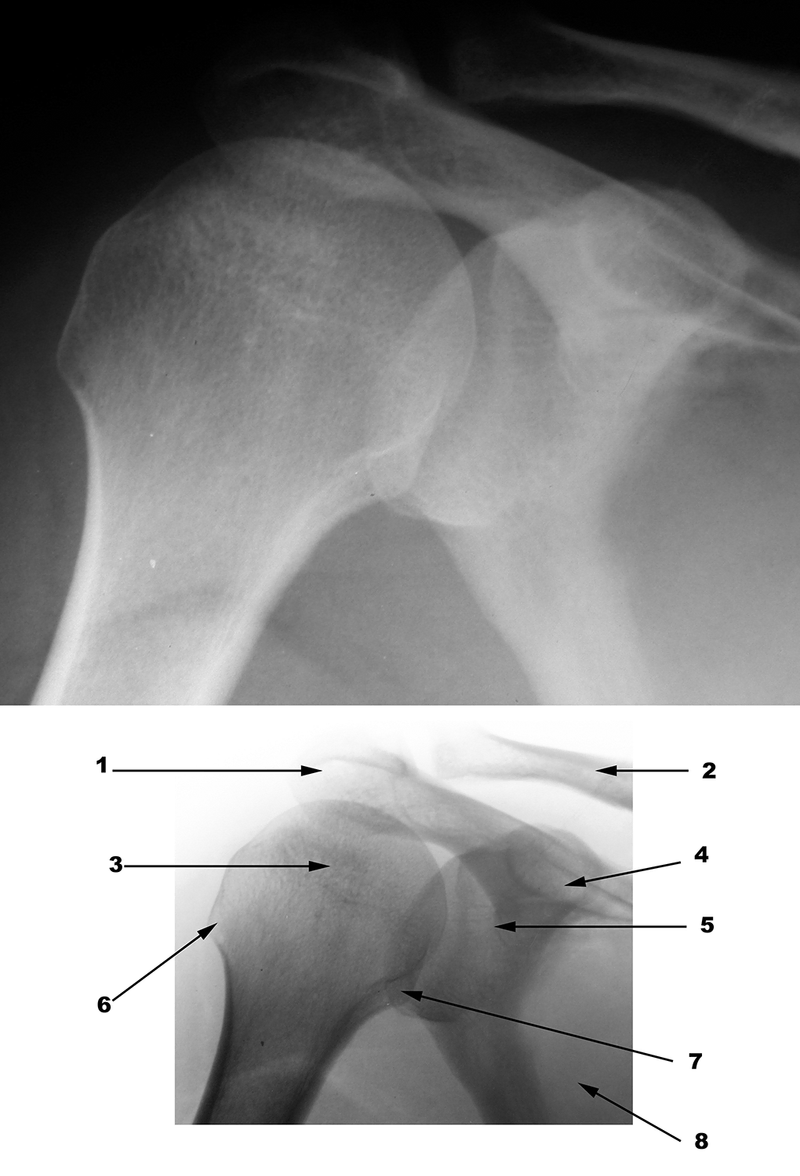
Regional Radiographic Anatomy:
The chapter explores radiographic anatomy by region, providing a detailed look at how different parts of the body appear in X-rays. From the shoulder and arm to the foot and ankle, each region has its own unique radiographic characteristics. The chapter also covers the different views and techniques used to capture the best possible images of each area.
Common Pathologies:
The final section of the chapter delves into common musculoskeletal conditions that are clearly visible on radiographs. Fractures, dislocations, arthritis, and tumors are just some of the pathologies that can be identified using this powerful imaging technique.
The Takeaway:
Radiography is a vital tool in modern medicine, providing a non-invasive way to visualize the inner workings of the human body. While it has its limitations, it remains an essential diagnostic technique for a wide range of musculoskeletal conditions. So, the next time you or a loved one needs an X-ray, remember the fascinating science behind this everyday medical procedure!