This chapter discusses synovial tissue, which is found in joints, tendon sheaths, and bursae and facilitates smooth motion. The synovium, a membranous structure, lines these areas and produces synovial fluid, which is crucial for motion and metabolism. The chapter covers the physiology, histology, and anatomy of normal synovium and synovial fluid, and then explores various pathologic conditions that can affect the synovium.
The physiologic function of the synovium is to secrete fluid, allowing smooth passage of tendons and decreasing friction in joints, tendon sheaths, and bursae. Synovial joints have opposing surfaces covered with articular cartilage and are enclosed by a capsule lined with synovium. This structure creates a pouch containing synovial fluid. The synovium also surrounds intra-articular ligaments and tendons.
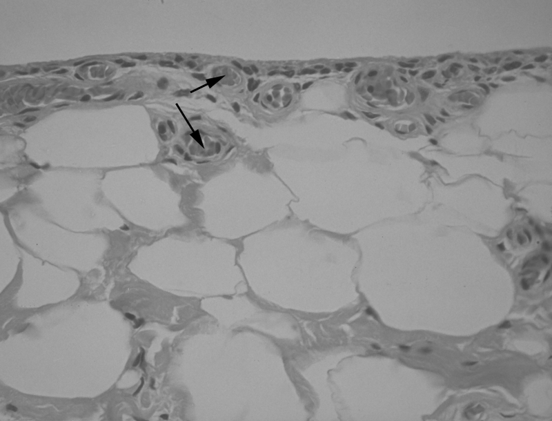
Histologically, the synovium consists of type A synoviocytes (macrophage-like) and type B synoviocytes (fibroblastic). Type A cells remove debris, while type B cells secrete hyaluronic acid. Grossly, the synovium is two to four cell layers thick and has a large surface area due to villi and microvilli. It is supported by a rich vascular network and other cells.
Synovial fluid is a clear, straw-colored fluid formed by the filtration of capillary plasma. It contains electrolytes and glucose in similar concentrations to plasma, but has lower concentrations of large molecules and proteins. The fluid's viscosity is dependent on the concentration of hyaluronic acid.
The chapter also discusses various conditions affecting the synovium. Synovitis, or inflammation of the synovial lining, can be caused by inflammatory or noninflammatory joint diseases. Examples of inflammatory diseases include bacterial septic arthritis and rheumatoid arthritis (RA). Noninflammatory diseases include osteoarthritis (OA). The chapter also details effusions, which are collections of synovial fluid that occur in both inflammatory and noninflammatory states.
Finally, the chapter touches on benign synovial tumors such as pigmented villonodular synovitis (PVNS) and primary synovial chondromatosis, and ongoing research areas like synovial supplementation and the etiology of RA.