This chapter discusses articular cartilage, the tissue that forms the surfaces of synovial joints. It highlights the structure, composition, and function of articular cartilage, emphasizing its role in enabling smooth joint movement and absorbing mechanical loads.Key points include:
- Composition: Articular cartilage is composed of an extracellular matrix (ECM) containing water, proteoglycans, and collagens, with chondrocytes distributed throughout.
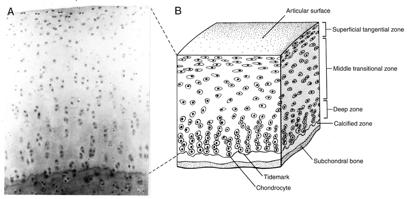
- Structure: The cartilage is divided into four zones: superficial tangential, middle (transitional), deep, and calcified, each with distinct cell shapes, collagen orientation, and proteoglycan concentration. The ECM is further divided into pericellular, territorial, and interterritorial regions.
- Function: Articular cartilage provides a low-friction surface for joint movement, absorbs mechanical load, and distributes it to the subchondral bone.
- Metabolism: Chondrocytes maintain the ECM by synthesizing and secreting its components, as well as regulating degradative enzymes.
- Response to Loading: Joint motion and loading are essential for maintaining cartilage health. Abnormal loading can lead to cartilage degeneration.
- Injury and Repair: Superficial cartilage injuries typically do not heal, while injuries that extend to the subchondral bone can initiate a repair response, though the repair tissue is often fibrocartilage, not hyaline cartilage.
- Osteoarthritis (OA): OA involves the progressive loss of articular cartilage, accompanied by attempted repair, bone remodeling, and osteophyte formation.
- Research and New Directions: Current research focuses on the relationship between chondrocytes and the ECM, mechanical signal transduction, growth factors, transplantation, and medications to inhibit matrix degradation.
In essence, the chapter provides a comprehensive overview of articular cartilage, from its basic biology to its pathology and potential avenues for future treatment.